By: Sridevi R. Pitta, MD, FSCAI, Rajiv Gulati, MD, FSCAI and Verghese Mathew, MD, FSCAI
Transfemoral catheterization is still a common procedure worldwide and vascular access bleeding during coronary interventions is associated with adverse outcomes. An important strategy for reducing access site bleeding is to achieve the optimal location for femoral access. However, there is a paucity of data on how well this goal is actually achieved in clinical practice using the traditional approaches of physical and fluoroscopic landmarks and micropuncture techniques.1
Location of Femoral Artery Access and Correlation with Vascular Complications
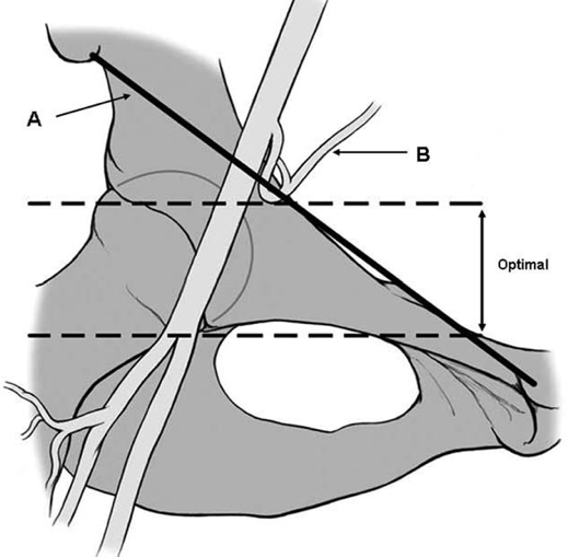
Arterial access above the femoral bifurcation but below the inferior border of the inferior epigastric artery is the optimal location and those that are either above or below these landmarks are suboptimal locations. In a 2011 study, the femoral artery access site was located outside the optimal location in 38 (13.0%) patients, and was associated with an increased risk of vascular complications. Overall, access related complications occurred in 17 (5.7%) patients and vascular complications were significantly more frequent in patients who had a femoral artery access outside the optimal location (18% vs. 4%, p< 0.001).1
Figure 1: Femoral Artery Anatomy
 |
The incidence of major femoral bleeding declined significantly from the earliest to the contemporary time period in a Mayo Clinic series of 17,000 patients.2 As noted in this series, adverse outcomes of major femoral bleeding included prolonged hospital stay and increased requirements for blood transfusion. Blood transfusion and major femoral bleeding were both associated with decreased long-term survival, driven by a significant increase in 30-day mortality. This should be a strong clinical driver for further improvements in access strategies for patients undergoing percutaneous coronary intervention (PCI). 2
Real Time Ultrasound Guidance Facilitates Femoral Arterial Access and Reduces Vascular Complications
Ultrasound guidance is widely used for central venous access in numerous hospital settings such as emergency departments (ED), intensive care units (ICU) and radiology, but is seldom used in the cardiac catheterization lab (Cath lab), despite growing evidence of its potential benefit and utility. Seto et al have shown that ultrasound (US) guidance compared to traditional landmark and fluoroscopic technique has reduced the number of necessary attempts for successful common femoral artery (CFA) cannulation, the time to vascular access, the risk of venipunctures, and subsequent vascular complications.3 Increasing US experience was associated with a reduced time required for access with US guidance; operators with more than 10 procedures have reduced access time and demonstrate a trend toward improved CFA cannulation success. Whichever access strategy is utilized in the Cath lab, there is growing evidence to show that US-guided vascular access is safe and effective.4
Ultrasound Guidance Access: Safe and Efficient
Ultrasound machines are increasingly available in numerous hospital settings, including medical floors, ICUs and EDs, and have been widely used by vascular surgeons, interventional radiologists, ER physicians, and ICU physicians for their vascular access procedures.5, 6 Only in the Cath lab has the use of ultrasound been notably limited, despite the significant potential for bleeding and other vascular complications from coronary, peripheral, and structural interventions. It is a straightforward technique that is relatively easy to learn and utilizes equipment that is readily available in most hospitals. With reasonable experience, US guidance facilitates precise cannulation of vessels regardless of anatomic variation, which can increase procedural success. In comparative studies of palpation-guided and US-guided radial access the number of needed attempts was reduced, the first-pass success rate was improved, and the time to access was decreased.7, 8, 9, 10 Not only is there increasing evidence to support US guidance for vascular access for coronary interventions, there are multiple additional applications of ultrasound in the cardiac catheterization lab. Use of US guided peripheral interventions has shown improved success rates in limb salvage and amputation prevention procedures.11, 12, 13, 14 Point-of-care US is affordable and easy to use in assisting in accurate vascular access. There are several ultrasound devices commercially available for vascular access.
A reference list is found below, in addition to comprehensive slide presentations with embedded video clips demonstrating various aspects of the techniques of US guided vascular access.
References:
- Pitta SR, Prasad A, Kumar G, Lennon R, Rihal CS, Holmes DR. Location of femoral artery access and correlation with vascular complications. Catheter Cardiovasc Interv. 2011.
- Doyle BJ, Ting HH, Bell MR, Lennon RJ, Mathew V, Singh M, Holmes DR, Rihal CS. Major femoral bleeding complications after percutaneous coronary intervention: incidence, predictors, and impact on long-term survival among 17,901 patients treated at the Mayo Clinic from 1994 to 2005. JACC Cardiovasc Interv. 2008.
- Seto AH, Abu-Fadel MS, Sparling JM, et al. Real-time ultrasound guidance facilitates femoral arterial access and reduces vascular complications: FAUST (Femoral Arterial Access with Ultrasound Trial). JACC Cardiovasc Interv. 2010; 3:751-758.
- Sobolev M, Slovut DP, Lee Chang A, Shiloh AL, Eisen LA Ultrasound-Guided Catheterization of the Femoral Artery: A Systematic Review and Meta-Analysis of Randomized Controlled Trials. JACC Cardiovasc Interv. 2010.
- Guidance on the use of ultrasound locating devices for placing central venous catheters. National Institute for Clinical Excellence. London (UK) September 2002. ISBN: 1-84257-213-X.
- Making Health Care Safer: A Critical Analysis of Patient Safety Practices. Chapter 21: Ultrasound Guidance of Central Vein Catheterization. Agency for Healthcare Research and Quality (USA) Evidence Report/Technology Assessment No.43, AHRQ Publication 01-E058. July 20, 2001.
- Rafie IM, Uddin MM, Ossei-Gerning N, Anderson RA, Kinnaird TD. Patients undergoing PCI from the femoral route by default radial operators are at high risk of vascular access-site complications. EuroIntervention. 2014.
- Jolly SS, Yusuf S, Cairns J, et al; RIVAL trial group. Radial versus femoral access for coronary angiography and intervention in patients with acute coronary syndromes (RIVAL): a randomized, parallel group, multicenter trial. Lancet. 2011; 377:1409-1420.
- Seto AH, Roberts JS, Abu-Fadel MS, Czak SJ, Latif F, Jain SP, Raza JA, Mangla A, Panagopoulos G, Patel PM, Kern MJ, Lasic Z. Real-time ultrasound guidance facilitates transradial access: RAUST (Radial Artery access with Ultrasound Trial). Crit Care. 2014.
- Shiloh A. Ultrasound-guided catheterization of the radial artery: a systematic review and metanalysis of randomized controlled trials. Chest. 2011; 139:524-529.
- Yeow KM, Toh CH, Wu CH, et al. Sonographically guided antegrade common femoral artery access. J Ultrasound Med. 2002; 21:1413-1416.
- Kweon M, Bhamidipaty V, Holden A, Hill AA. Antegrade superficial femoral artery versus common femoral artery punctures for infrainguinal occlusive disease. J Vasc Interv Radiol. 2012; 23:1160-1164.
- Yilmaz S, Sindel T, Lüleci E. Ultrasound-guided retrograde popliteal artery catheterization: experience in 174 consecutive patients. J Endovasc Ther. 2005; 12:714-722.
- Arnold Seto, MD, MPA, FACC, FSCAI, and Mazen Abu-Fadel, MD, FACC, FSCAI Ultrasound-Guided Arterial Access Is the Way to Go. Vascular access using ultrasound is safe and efficient. Cardiac Interventions Today. 2012.
Related QI Tips
Other evidence-based methods and tools you can use to improve quality of care and outcomes for patients.
