Dean Ferrera, DO, FSCAI; Jaafer Golzar, MD, FSCAI; and Faisal Latif, MD, FSCAI
Interventional cardiologists commonly employ revascularization techniques to assist in the care of aortoiliac, infrainguinal, and infrapopliteal obstructive peripheral artery disease (PAD). Direct percutaneous access of the pedal vasculature is an essential skill set for many endovascular physicians, and the adjunctive use of retrograde pedal access has been demonstrated to assist in recrossing complex infrapopliteal lesions.1 Moreover, use of pedal access has been shown to improve crossing success after previously failed attempts at lesion crossing, especially in the setting of critical limb ischemia.1, 2, 3, 4 Through careful angiographic and ultrasound guidance, access of the anterior tibialis, posterior tibialis, and peroneal arteries remains feasible and safe.5
This Tip of the Month will focus on specifically choosing the best pedal access site to help achieve revascularization of target vessels, including cannulation points. Also, commonly used devices and techniques for transpedal interventions will be reviewed, and a case example will be provided to highlight some options when working from the pedal access sites.
Choosing the Pedal Access Site
In general, the choice of pedal artery access site is best determined by the intended target vessel to be treated. Retrograde access can be used to cross infrapopliteal, popliteal, and superficial femoral artery occlusive disease. Retrograde access can be considered in any segment of the vessel, although more proximal access requires a greater degree of expertise. Given the deep position of the peroneal artery in the posterior compartment of the lower leg, access of this vessel is reserved only for inaccessible anterior tibialis (AT) and posterior tibialis (PT) vessels. The more superficial positions of the AT and PT in the distal lower limb also lend easier hemostasis and control of the access site post-procedure.
Working From the Pedal Access Site
Achieving success from the pedal access site includes a good working knowledge of facilitating equipment. In general, the pedal vessels can accommodate 0.14”, 0.18”, and 0.35”catheters and support wires. Access is typically achieved with 21g needles and 0.18” wires. Bareback access is possible in order to maintain a low profile; however, a sheath can safely be placed in the pedal vessels if necessary. Hydrophilic catheters and sheaths are preferred to mitigate arterial spasm. Slender® sheaths by Terumo (Tokyo, Japan) are often advantageous due to their ability to offer internal diameters of up to 6Fr on an outer diameter size of 5Fr. Pedal access kits are available and offer a micropuncture catheter and needle for access with a check-flow cap. These catheters offer the smallest footprint in the pedal position with an outer diameter of 4Fr and a smaller working internal diameter of 2.9Fr.
Dual access from the contralateral limb or antegrade femoral access is typically helpful for angiographic guidance, aiding in visualization of the pedal vessels and allowing for the externalization of the retrograde wire via snare or direct sheath/catheter wiring techniques. Working completely pedal is sometimes preferred though.
For such purpose, a useful catheter series often includes the following: TrailBlazer™ series by Medtronic (Minneapolis, MN), CXI® series by Cook, Angled GLIDECATH® by Terumo, or an internal mammary catheter.
After crossing the lesion in question, it is usually helpful to snare the retrograde wire, externalize it, and proceed to work in an antegrade approach. Alternatively, angioplasty can often be performed from the pedal position, with then an attempt to pass an antegrade wire target past the lesion. When there is a desire to perform atherectomy, small working devices such as a 1.25mm Micro Diamondback® by Cardiovascular Systems, Inc. (Minneapolis, MN) or small laser fibers of 0.9” to 1.4” can often be deliverable to the target lesion. Ultimately, when choosing atherectomy devices, consideration must be given to not only the target vessel size, but also the size of the access site sheath.
Table 1: Pedal access sites for peripheral intervention
|
Anterior Tibialis |
Posterior Tibialis |
Peroneal |
|
|
Ultrasound Visualization |
Distal anterior compartment |
Distal posterior compartment |
Distal deep posterior compartment |
|
Sheath Size |
Bare catheter, 4F; 5F |
Bare catheter, 4F; 5F |
Bare catheter, 4F; 5F |
|
Position |
Superficial in distal limb |
Superficial in distal limb |
Deep, posterior to tibia |
|
Typical Target Vessels |
AT; Pop; SFA |
PT; TPT; Pop; SFA |
P; TPT; Pop; SFA |
Abbreviations: AT – anterior tibialis; P – peroneal; Pop – popliteal artery; PT – posterior tibialis; SFA – superficial femoral artery; TPT – tibioperoneal trunk.
Case Example
History:
- The patient is a 67-year-old male with Rutherford class 4 critical limb ischemia (CLI).
- There is poor healing at prior digit amputation sites 1 and 2.
- The patient has a history of diabetes mellitus, coronary artery disease (CAD), former smoking, and obesity.
- A diagnostic peripheral angiography demonstrated severe L SFA disease, previously treated with directional atherectomy and drug-coated balloon angioplasty.
- The patient has known below-the-knee PAD.
Technique:
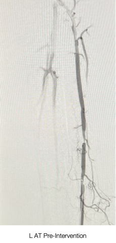
- Ultrasound-guided access of the distal AT above the ankle was performed, and a 5Fr Slender® sheath was placed.
- The AT lesion was crossed using a Choice™ PT floppy wire (Boston Scientific, Marlborough, MA).
- A straight 0.18” CXI® catheter was used to exchange wires proximal to the stenosis for a CSI Viper®
- An orbital atherectomy was performed with a CSI Diamondback®25mm Micro Crown at low and medium speeds.
- A balloon angioplasty of the AT lesion was performed with a NanoCross®0mm x 100mm balloon.
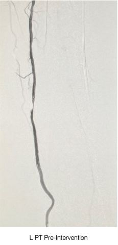
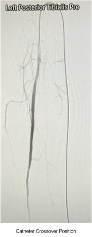
- Crossover to the L PT was performed using an angled 0.35” TrailBlazer® support catheter and Choice™ PT floppy wire.
- Orbital atherectomy was performed with the same CSI Dia
- Hemostasis was achieved at the end of the procedure with sheath removal and a Terumo TR Band®.
Conclusions
- Utilization of pedal access sites can aid in lesion crossing via an inline direct access point, particularly in patients with CLI and a previously failed intervention using antegrade access.
- With careful procedural planning, infrainguinal atherectomy and angioplasty procedures can be readily performed.
- Use of ultrasound attention to lower-extremity anatomical principles and a good working knowledge base of facilitating equipment will all help the operator find success in performing peripheral interventions from the pedal access position.
References
- Montero-Baker M, Schmidt A, Braünlich S, et al. Retrograde approach for complex popliteal and tibioperoneal occlusions. J Endovasc Ther. 2008 Oct;15(5):594-604.
- Bazan HA, Le L, Donovan M, et al. Retrograde pedal access for patients with critical limb ischemia. J Vasc Surg. 2014 Aug;60(2):375–81.
- Walker C. Pedal access in critical limb ischemia. J Cardiovascular Surg (Torino). 2014 Apr; 55(2):225–7.
- Taha AG, Abou Ali AN, Al-Khoury G, et al. Outcomes of infrageniculate retrograde versus transfemoral access for endovascular intervention for chronic lower extremity ischemia. J Vasc Surg. 2018 Oct;68(4):1088–1095.
- Mustapha JA, Saab F, McGoff T, et al. Tibio-pedal arterial minimally invasive retrograde revascularization in patients with advanced peripheral vascular disease: the TAMI technique, original case series. Catheter Cardiovasc Interv. 2014 May 1;83(6):987–94.
Related QI Tips
Other evidence-based methods and tools you can use to improve quality of care and outcomes for patients.